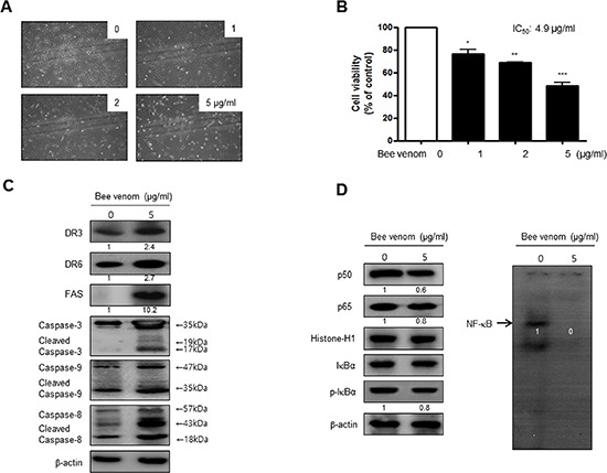
Figure 3. Effect of BV on cell viability and morphological changes of human primary cervical cancer cells.

Morphologic observation with the treatment of BV. Human primary cervical cancer cell changes were observed under phase contrast microscope (A). The data (B) are expressed as the mean ± S.D. of three experiments. *(P < 0.05) indicates statistically concentration-dependent effect of BV (A) on the MTT viability assay in human primary cervical cancer cell. Expression of apoptosis regulatory proteins related extrinsic pathway was determined using Western blot analysis (C), NF-κB activity and expression of related proteins determined by EMSA or Western blot (D) were determined as similar to the cancer cell lines. Each band is representative for three experiments.
