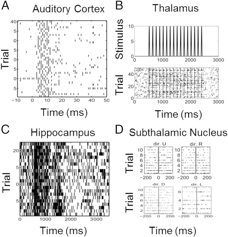
Fig. 1.
Raster plots of neural spiking activity. (A) Forty trials of spiking activity recorded from a neuron in the primary auditory cortex of an anesthetized guinea pig in response to a 200 μs/phase biphasic electrical pulse applied in the inferior colliculus at time 0. (B) Fifty trials of spiking activity from a rat thalamic neuron recorded in response to a 50 mm/s whisker deflection repeated eight times per second. (C) Twenty-five trials of spiking activity from a monkey hippocampal neuron recorded while executing a location scene association task. (D) Forty trials of spiking activity recorded from a subthalamic nucleus neuron in a Parkinson's disease patient before and after a hand movement in each of four directions (dir.): up (dir. U), right (dir. R), down (dir. D), and left (dir. L).

