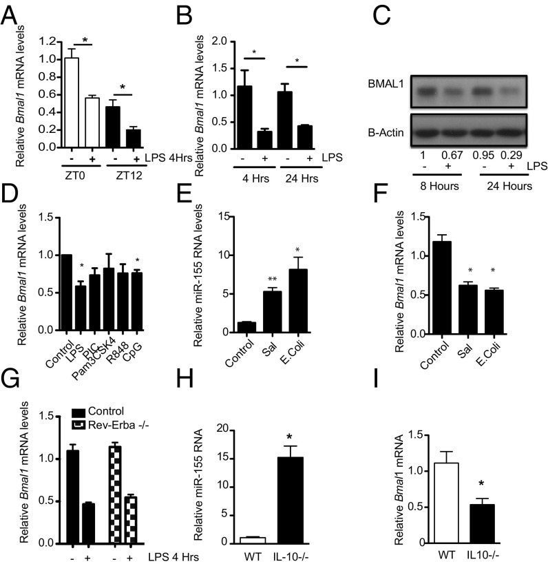
Fig. 2.
Activation of macrophages represses the core clock gene Bmal1, coincident with up-regulation of MiR-155. (A) Wild-type peritoneal cells harvested at ZT0 and ZT12 and treated immediately ex vivo with LPS (100 ng/mL) for 4 h and analyzed for Bmal1 (n = 3). (B) BMDMs were exposed to LPS (100 ng/mL) for the indicated times and analyzed for Bmal1 (n = 4–5). (C) BMAL1 protein by immunoblot (representative of n = 3). Values provided are relative band intensity corrected for β-actin. (D) BMDMs were treated with LPS (100 ng/mL), Poly-I:C (PIC, 100 µg/mL), Pam3CSK4 (100 ng/mL), R848 (1 µg/mL). and CpG (1 µg/mL) for 4 h and analyzed for Bmal1 (n = 3). RAW-264 cells exposed to E. coli or Salmonella for 24 h and analyzed for expression of (E) miR-155 and (F) Bmal1 (n = 3). (G) BMDMs from wild-type and Rev-Erbα −/− mice were treated with LPS (100 ng/mL) for 4 h and analyzed for Bmal1 (n = 3). BMDMs harvested from control and Il-10−/− mice and analyzed for expression of (H) miR-155 and (I) Bmal1 (n = 8–10). *P ≤ 0.05 and **P ≤ 0.01.

