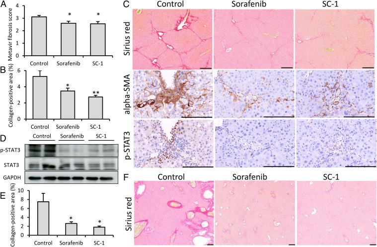
Fig. 1.
Sorafenib and SC-1 treatment ameliorate liver fibrosis in hepatotoxic and cholestatic mouse models. In the thioacetamide-induced liver fibrosis mouse model (n = 9–10 in each group), significant fibrosis regression was found after sorafenib and SC-1 treatment by Metavir fibrosis scoring (A) and collagen-positive area quantification (B). (C) Representative liver tissues after Sirius Red, α-SMA, and p-STAT3 immunohistochemical staining. Significant fibrosis regression after sorafenib and SC-1 treatment was observed as indicated by Sirius Red staining. α-SMA and p-STAT3 overexpression was reduced after sorafenib and SC-1 treatment. (D) Hepatic p-STAT3 was down-regulated after sorafenib and SC-1 treatment. In the bile duct ligation mouse model (n = 4–6 in each group), significant fibrosis regression was observed after sorafenib and SC-1 treatment by collagen-positive area quantification (E) and Sirius Red staining (F). Columns, mean; bars, SE. *P < 0.05, **P < 0.01. (Scale bars: 200 μm.)

