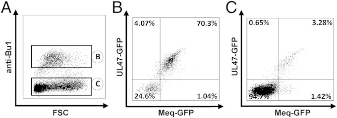
Fig. 3.
Transfer of MDV from B to T cells. (A) B cells were infected with RB1B-UL47-RFP_Meq-GFP for 24 h, infected B cells sorted by FACS and cocultured with TCR-2–stimulated thymic T cells for 2 d. Cultures were stained with anti-chBu1 to discriminate between B and non-B thymocytes (predominantly T cells). Analysis of B cells (B) and non-B cells (C) in the culture. Percentage of infected B cells is shown from one representative experiment.

