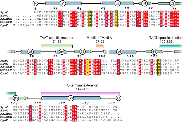
Fig. 2.
Multiple sequence alignment for key members of the TAAT family. Secondary structure of ApxC (consensus of the four monomers in the crystal structure) is shown immediately above the alignment of TAAT sequences ApxC (A. pleuropneumoniae), HlyC (Uropathogenic E. coli), AaLktC (A. actinomycetemcomitans), MhLktC (M. hemolytica), and CyaC (Bordetella pertussis). The α-helices indicated are shown as tubes (blue); β-strands by arrows (green). Conserved residues are highlighted red, and important active site residues identified later (His24, Asn35, Asp93, and Arg121) are shown in gold. Features that differentiate TAATs from other GNATs are highlighted by using the same color scheme as in Fig. 1B.

