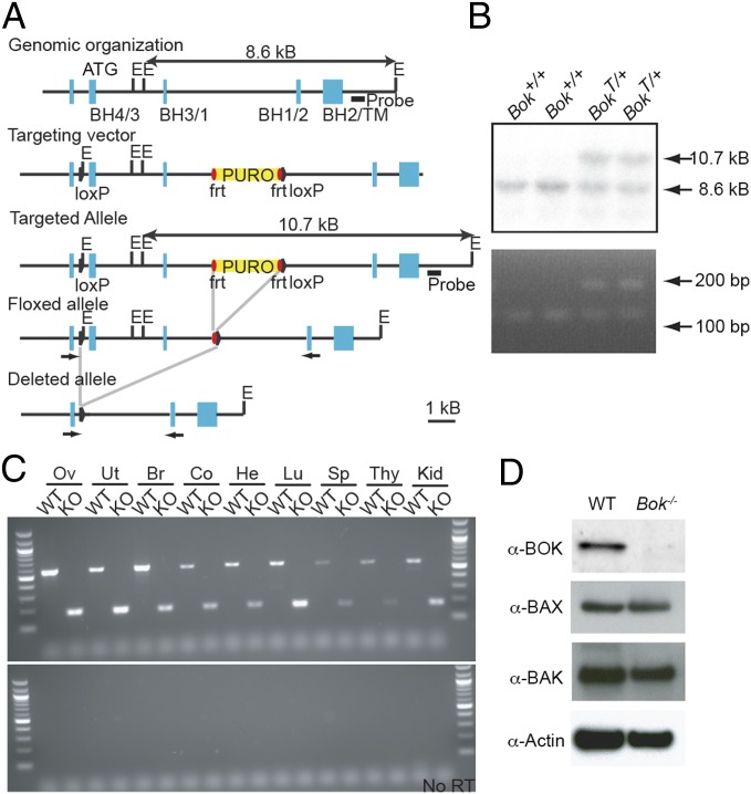
Fig. 1.
Targeted disruption of the mouse Bok gene. (A) Partial map of the murine Bok locus (Top), Bok targeting vector (Top Middle), targeted homologous recombination (Middle), removal of the puromycin resistance (puro) cassette (Bottom Middle), and complete deletion of the locus (Bottom). Bok exons are shown as blue boxes with positions of BCL-2 homology (BH) and transmembrane (TM) domains indicated below. Solid triangles represent LoxP sites, and red ovals indicate FRT sites. The probe used in Southern blotting is shown as a black line. Arrows represent primers used for PCR genotyping in B and RT-PCR in C. E, EcoRI. (B) Southern blot analysis of ES cell DNA showing the presence of WT (+/+) and heterozygous (+/Targeted) mutant genotypes (Top). PCR analysis of ES cell DNA showing the presence of the 5′ LoxP site in targeted clones (Bottom). (C) RT-PCR of Bok in multiple tissues from WT and Bok−/− mice: ovary (Ov), uterus (Ut), brain (Br), colon (Co), heart (He), lung (Lu), spleen (Sp), thymus (Thy), and kidney (Kid). (D) Western blot for BOK, BAX, BAK, and Actin from WT and Bok−/− MEFs.

