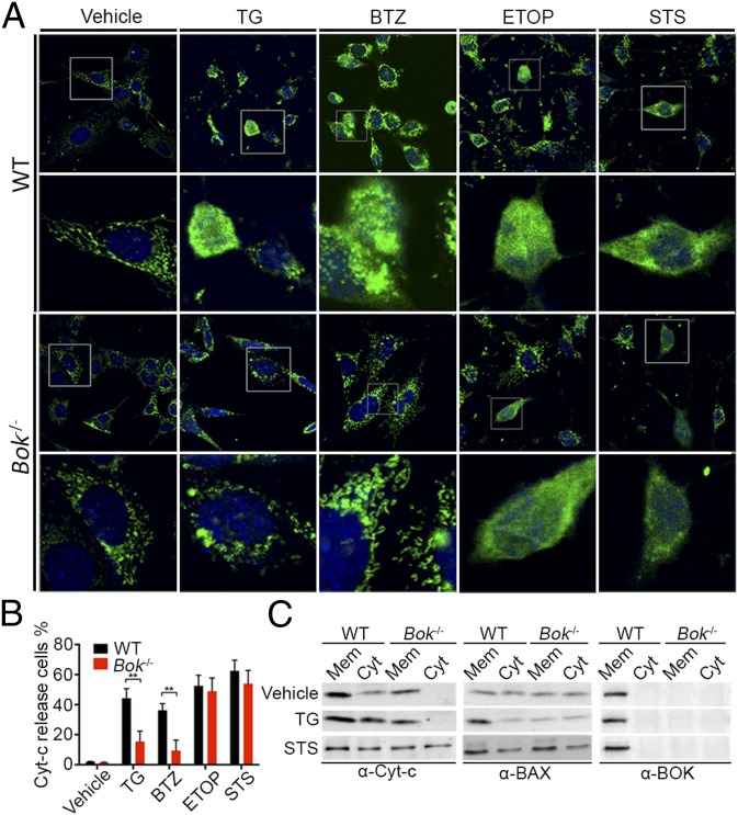
Fig. 4.
BOK promotes cytochrome c release and BAX translocation in response to ER stress. (A) Immunofluorescence detection of cytochrome c release by SV40-immortalized WT and Bok−/− cells subjected to vehicle or 24-h exposure to 10 μM TG, 10 μM BTZ, 10 μM ETOP, or 1 μM STS. Cytochrome c release and cell nuclei were examined by confocal immunofluorescence microscopy using anti-cytochrome c antibody (green) and TO-PRO3 (blue) staining. (B) Quantitative analysis of cytochrome c release from A. For each condition, 100 cells were analyzed in each of five randomly chosen fields. Three independent experiments were performed, and the means ± SEM were plotted. Significance was calculated using an ANOVA test (**P < 0.005). (C) Representative Western analyses of cytosolic (Cyt) and membrane (Mem) fractions of WT and Bok−/− cells subjected to vehicle or 24-h treatment with 10 μM TG or 1 μM STS to monitor cytochrome c release (Left), and BAX (Middle) and BOK (Right) translocation. The presence of mitochondria and ER in the membrane preparation is further documented by Western analyses for COX IV and calnexin, respectively (Fig. S4).

