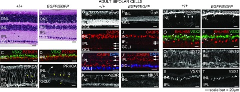
Fig. 4.
RB and type 2 OFF-cone BP cells are significantly reduced in the adult PRDM8-null retina. (A and B) H&E-stained retinal sections from adult WT and Prdm8EGFP/EGFP mice showed that Prdm8EGFP/EGFP mice have abnormally thin INL and IPL layers. (C–T) Representative confocal micrographs of retinal cryosections from adult Prdm8EGFP/EGFP mice and WT littermates (8–12 wk old) stained for RB and CB cell markers. (C and D) BP cell (VSX2+P27KIP1−) numbers were reduced in Prdm8EGFP/EGFP retina compared with WT, whereas numbers of Müller glia (VSX2+P27KIP1+ orange cells) were unchanged. (E and F) PRKCA expression in RB cells was nearly absent from the Prdm8EGFP/EGFP retina, but (F) a few PRKCA+ cells showing RB cell-like morphology persisted (white arrowhead), whereas the number of PRKCA+ ACs was significantly increased (yellow arrowheads). (G and H) Goα ON-BP staining of the INL and IPL was greatly reduced overall, with little staining in the INL and strongly diminished staining in the IPL. (I–J′) CABP5 staining (RB and types 3 and 5 CB cells) was reduced in Prdm8EGFP/EGFP retina. Furthermore, CABP5+ RB axonal projections to the innermost IPL were absent, such that only two bands of staining in the IPL were distinguishable in Prdm8EGFP/EGFP retina, whereas three were visible in the WT (arrows); the lowest band of axon terminals abutting the GCL in (I and I′) WT retina was absent in (J and J′) mutants. (I′ and J′) Enlargement of areas from I and J showing GCL nuclei counterstained with DAPI (blue). (K and L) Types 1 and 2 CB marker NK3R was dramatically reduced in the Prdm8EGFP/EGFP retina vs. WT. (M–P) The type 2 CB markers, recoverin and BHLHB5, were significantly reduced among BP cells in the Prdm8EGFP/EGFP retina. (O and P) Although there were fewer BHLHB5+VSX2+ BP cells in Prdm8EGFP/EGFP retina (arrowheads), the number of BHLHB5+VSX2− ACs was unchanged. (Q–T) The numbers of SYT2+ cells (types 2 and 6 CB cells) and VSX1+ nuclei (types 1, 2, and 7 CB cells) were reduced in the Prdm8EGFP/EGFP retina vs. WT. ONL, outer nuclear layer.

