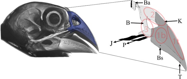
Fig 1. Schematic representation of our multi-layered (bone: B, keratin: K) finite element modeling approach, for the medium ground finch Geospiza fortis.

Bending area (Ba) and bite position (base: Bs, or tip: T) were constrained in our models for translation and rotation, and muscle forces were applied in our models via the jugal (J) and palatine (P) jaw bones (black elements are constrained). Locations of vM Stress recordings are indicated with transparant ellipses (1b: on top of bone, near base bite position; 1t: on top of bone near tip bite position; 2: on top of the beak near the nasal hinge; 3: nasal bone).
