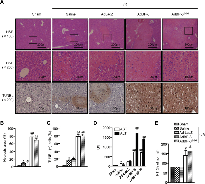
Figure 2. Aggravation of hepatic I/R injury by IGFBP-3.
(A) After 24 h reperfusion, liver necrosis and apoptosis were assessed by H&E and TUNEL staining methods, respectively. (B) The area of necrosis was analyzed. (C) Apoptotic cells were counted and expressed as a percentage of all hepatocytes. (D) After 6 h reperfusion, serum levels of AST and ALT were analyzed. (E) After 24 h reperfusion, prothrombin time (PT) was analyzed. Values are the mean ± SEM (n = 9 mice per group). *, p < 0.05 versus sham-operated mice; #, p < 0.05 and ##, p < 0.01 versus AdLacZ-injected mice. AdBP-3, AdIGFBP-3; AdBP-3 GGG, AdIGFBP-3GGG.

