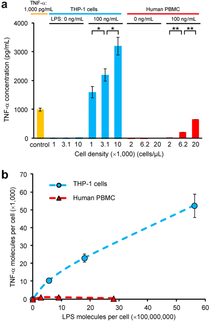
Figure 5. Detection of TNF-α secreted from THP-1 cells and human peripheral blood mononuclear cells (PBMCs) using the integrated microfluidic immunosensing chip.

(a) Plot of TNF-α concentration as a function of THP-1 cell and human PBMC density. Immune cells were either untreated (LPS: 0 ng mL−1) or pre-stimulated with LPS (100 ng mL−1), as indicated. A cell culture medium spiked with 1,000 pg mL−1 TNF-α was included as positive control for comparison. (b) Plot of average TNF-α molecules secreted by individual THP-1 cells and human PBMCs as a function of LPS concentration per cell. Data represents the mean ± S.D., and n = 3. P-values were calculated using unpaired student’s t-tests. *,P < 0.05 and **,P < 0.005.
