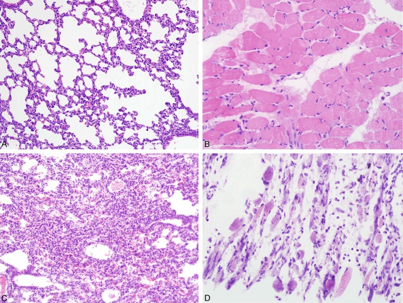
Figure 2.

Histological features of tissues of the infected mice-1. Pictures in the top row showed normal histological features of the lungs (A) and skeletal muscles (B) of mice in the control group. In EV71 infected mice, obvious inflammatory cells exudation was observed in alveolar spaces of lung tissues (C). The skeletal muscle tissues of the limbs presented myofibril fracture and myocyte disruption (D).
