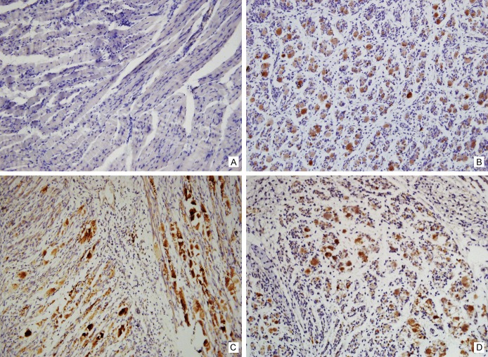
Figure 4.
Detection of VP1 protein of EV71 by immunohistochemistry method-1. Immunohistochemistry staining of the skeletal muscle tissues of the normal mice was as negative control (A). By contrast, the evident staining was exhibited in skeletal muscles of the EV71 infected mice, including the limbs (B), the intercostal spaces (C) and even along the spine (D).

