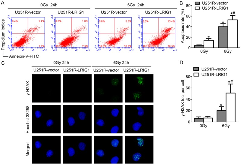
Figure 5.

Overexpression of LRIG1 enhances irradiation-induced apoptosis and DNA damage of U251R cells. (A)Representative plots of show Annexin-V/PI staining in U251R-vector and U251R-LRIG1 cells 24 h after treatment with or without ionizing radiation of 6 Gy. (B) The histogram represented quantitative analysis of the apoptotic rates as shown in (A). (C) Examples show residual immunofluorescence staining against γ-H2AX foci (green) and nucleus (blue); magnification, ×400. (D) Quantitative analysis of the number of γ-H2AX foci per cell. *P < 0.05 compared with U251R-vector cells without irradiation treatment. *#P < 0.05 compared with U251R-vector cells with irradiation treatment.
