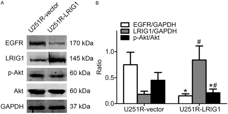
Figure 6.

LRIG1 regulates EGFR expression and Akt phosphorylation level. Equal amounts of cell protein were separated by SDS-PAGE and probed by anti-LRIG1, anti-EGFR, anti-Akt and anti-phosphorylated Akt antibody. GAPDH protein was used as loading control. A. Western blotting detection of proteins in U251R-vector and U251R-LRIG1 cells without treatment with ionizing radiation. B. The histogram represents quantitative analysis of EGFR, LRIG1, total and phosphorylated Akt. *P < 0.05 compared with U251R-vector cells; #P < 0.05 compared with U251R-vector cells; *#P < 0.05 compared with U251R-vector cells.
