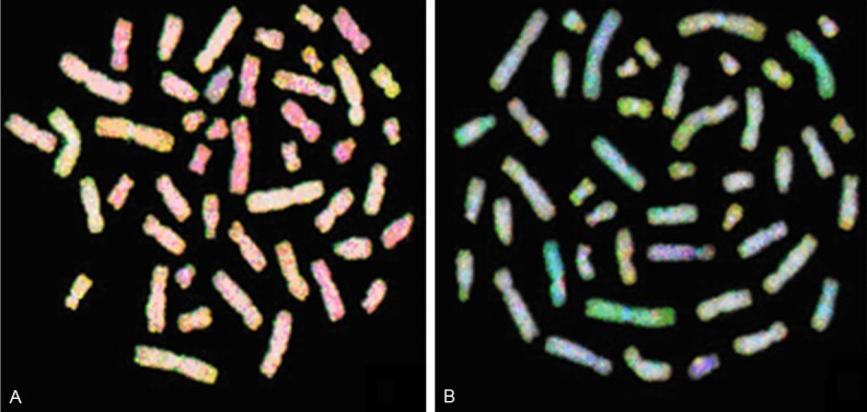
Figure 2.

Comparative genomic hybridization metaphase spreads of RCC 20 (A) and RCC 25 (B). Green areas are gains, red areas are losses, yellow/yellowish areas are normal, and blue areas are heterochromatin. Hybridization to repetitive sequences/heterochromatin were blocked by unlabeled human Cot-1 DNA and stained blue with 4,6-diamidino-2 phenylindole-2 HCL (DAPI).
