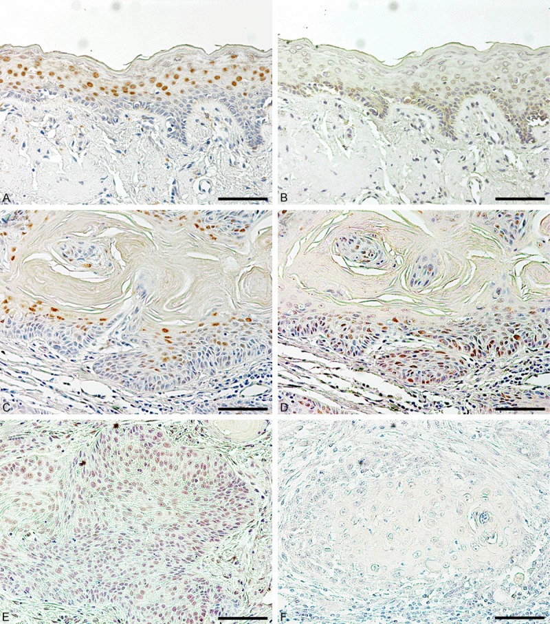
Figure 1.

Immunohistochemical staining of KLF4 and KLF5 in oral epithelium and carcinomas. Expression of KLF4 (A, C) and KLF5 (B, D, E) in normal oral epithelium adjacent to carcinoma cells (A, B) and in oral carcinoma tissues with (C, D) or without keratin pearls (E) was examined by immunostaining. Non-immune IgG instead of primary antibody was used as a negative control (F). Bar = 40 µm.
