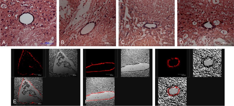
Figure 1.

HE staining of mild spinal cord injury at different time post-injury and Dil labeling the endogenous neural stem cells in ependymal regions 24 after injection. A. The histological morphology of normal spinal cord with HE staining. B. The histological changes of spinal cord tissue at 1d post-injury with HE staining. C. The histological changes of spinal cord tissue at 3d post-injury. D. The histological changes of spinal cord tissue at 7d post-injury. E. Dil labeling the lining ependymal cells of ventricular. F. Dil labeling the lining ependymal cells of the fourth ventricle. G. Dil labeling the cells of central canal of spinal cord at T10 level. Scar bars = 100 um in A-D, E and F = 40 um.
