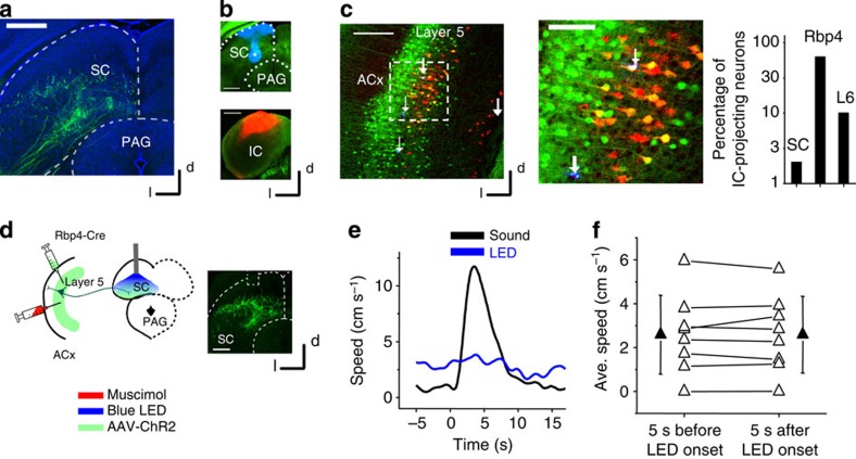
Figure 6. Corticofugal projections to SC unlikely contribute to the noise-induced flight behaviour.
(a) Labelling of corticofugal axons from L5 of the ACx to the intermediate and deep layers of SC. Scale bar, 500 μm. (b) Double injection of two different retrograde tracers into deep layers of SC (blue, top) and the ICx (red, bottom) of a Rbp4-Cre GFP mouse. Scale bar, 500 μm. Note that the blue staining in the superficial layer of SC was due to leakage (injection site marked by *). (c) Left, retrograde labelling of SC- and IC-projecting neurons in the ACx. Red neurons project to the IC, blue neurons (marked by white arrows) to the SC. SC-projecting neurons are scarce (only four neurons in the view field). Scale bar, 250 μm. Middle, a blow-up image of the boxed area on the left. One out of four SC-projecting neurons also projects to the IC (labelled by white colour). Scale bar, 100 μm. Right, percentage overlap between retrogradely labelled IC-projecting cortical neurons with retrogradely labelled SC-projecting cortical neurons, genetically labelled L5 cells in the Rpb4-Cre mouse and with layer 6 neurons. (d) Injection of AAV-ChR2-EYFP-abelled L5 axon terminals in SC deep layers. Blue LED was applied to the SC, while the ACx was silenced with muscimol. Right, fluorescence image showing the ACx-SC axon terminals and optic fibre placement. Scale bar, 500 μm. (e) Speed trace of an example animal in response to blue LED illumination of the SC (blue) and to 80 dB SPL noise (black). (f) Speeds averaged (Avg.) within a 5-s window before and after the onset of LED illumination in SC deep layers. N=8 animals. No difference was detected. P=0.97, two sided paired t-test. All error bars represent s.d.

