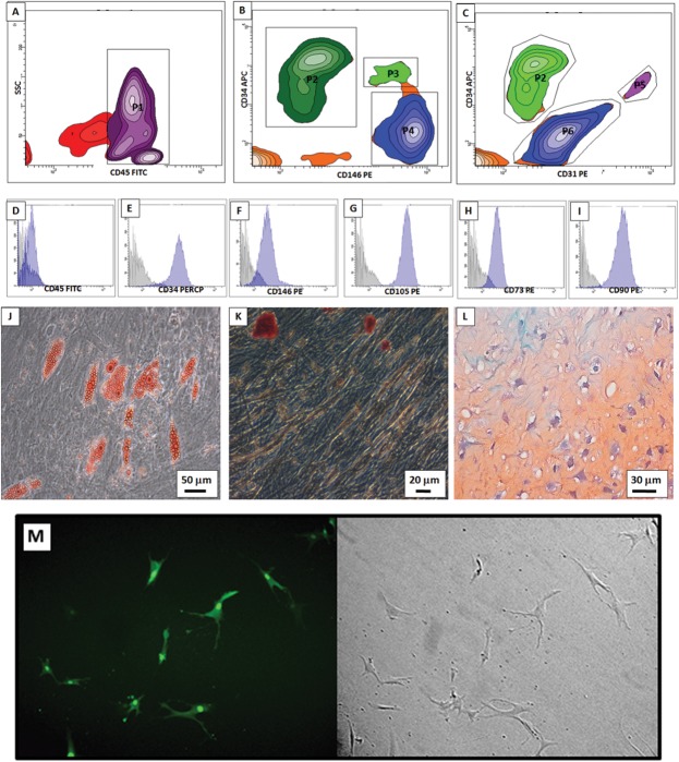
Fig 1. AT-MSC characterizations and Transduction efficiency.
In freshly isolated cells major two subpopulations were present: (A) Hematopoietic (P1—CD45 positive) and non-hematopoietic cells (events outside P1, CD45 negative). Non-hematopoietic cells were analyzed in (B) and (C). Pre-adipocytes were detected in B (P2—CD146 negative and CD34 positive) and in C (P2—CD31 negative and CD34 positive). MSC were detected in B (P5—CD146 positive, CD34 negative). Endothelial progenitors were detected in C (P5—CD31 positive, CD34 positive). (D—F) After seeding, AT-MSC from monolayer was still negative for CD45 and positive for CD34 and CD146. (G-I) AT-MSC were also positive for CD105, CD73 and CD90, which are all mesenchymal surface markers in vitro. (J—L) AT-MSC are multipotent for adipogenic (Oil Red O staining), osteogenic (Alizarin staining) and chondrogenic (Pellet culture, safranin staining) lineages, respectively. (M) Transduction efficiency in AT-MSC of the lentivirus containing Tk-GFP is 80%.

