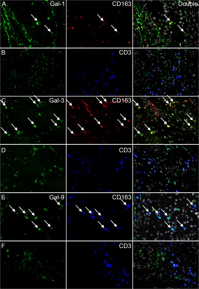
Fig 3. Immunofluorescent stainings of galectin-1, -3, -9, CD163 and CD3.
Representative images from double stainings of galectin-1 and CD163 (A) or CD3 (B), galectin-3 and CD163 (C) or CD3 (D) and galectin-9 and CD163 (E) or CD3 (F) are shown. Images containing both stainings and DAPI are shown in the right column. Arrows indicate examples of double positive cells.

