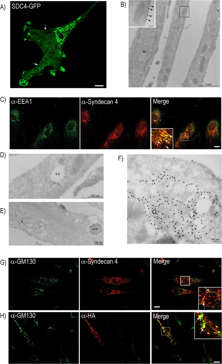
Fig 1. Syndecan-4 localization in bovine muscle cells.
A) Muscle cells transiently transfected with Syndecan-4 (SDC4)-GFP were fixed with 4% PFA before fluorescence microscopy analysis. Scale bar: 20 μm. Arrows show plasma membrane staining. B) Muscle cells transiently transfected with SDC4-HA were induced to differentiation before immuno-EM preparation. Thawed cryosections were labelled using anti-HA antibody and 15 nm protein A gold. Arrow heads in insert (higher magnification of framed area) indicate syndecan-4 staining at the plasma membrane. N: Nucleus. Scale bar in insert: 100 nm. C) Differentiating cells, fixed with 4% PFA, were immunostained with mouse anti-syndecan-4 and goat-anti EEA1 (both diluted in PierceImmunostain Enhancer), followed by Alexa 546-conjugated goat anti-mouse (red) and Alexa 647-conjugated donkey anti-goat (green) before fluorescence microscopy analysis. The insert represents high magnification of the framed area. Arrows denote syndecan-4 and EEA1 co-localization. Scale bar: 20 μm. D-F) Thawed cryosections of SDC4-HA transfected cells induced to differentiation were labelled using an anti-HA antibody and 15 nm protein A gold. Localization of SDC4-HA to plasma membrane and compartments with morphology resembling D) early endosomes (E.E.), E) multi vesicular bodies (MVB) and compartments with a reticular morphology (R), and F) the Golgi apparatus (G.). G) Differentiating cells, fixed with ice-cold ethanol, were immunostained with mouse anti-syndecan-4 (diluted in Pierce Immunostain Enhancer) and rabbit anti-GM130, followed by Alexa 488-conjugated goat-anti rabbit (green) and Alexa 546-conjugated goat-anti mouse (red) before fluorescence microscopy analysis. The insert represents high magnification of the framed area. Arrows indicate co-localization of the Golgi marker GM130 and syndecan-4. Scale bar: 20 μm. H) Muscle cells transfected with SDC4-HA and induced to differentiated were fixed with ice-cold ethanol and immunostained with mouse anti-HA and rabbit anti-GM130, followed by Alexa 488-conjugated goat-anti rabbit (green) and Alexa 546-conjugated goat-anti mouse (red) before fluorescence microscopy analysis. The inserts represents high magnification of the boxed area. Arrow indicates co-localization of the Golgi marker and anti-HA. Scale bar: 20 μm.

