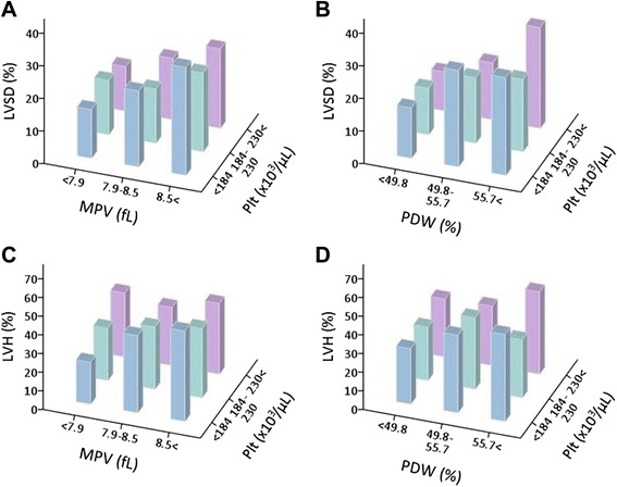
Fig. 3.

Prevalence of left ventricular systolic dysfunction (LVSD) and left ventricular hypertrophy (LVH) according to platelet indices. Shown is the prevalence of LVSD (a, b) and LVH (c, d) according to platelet count and mean platelet volume (MPV) tertiles (a, c), and platelet count and platelet distribution width (PDW) tertiles (b, d)
