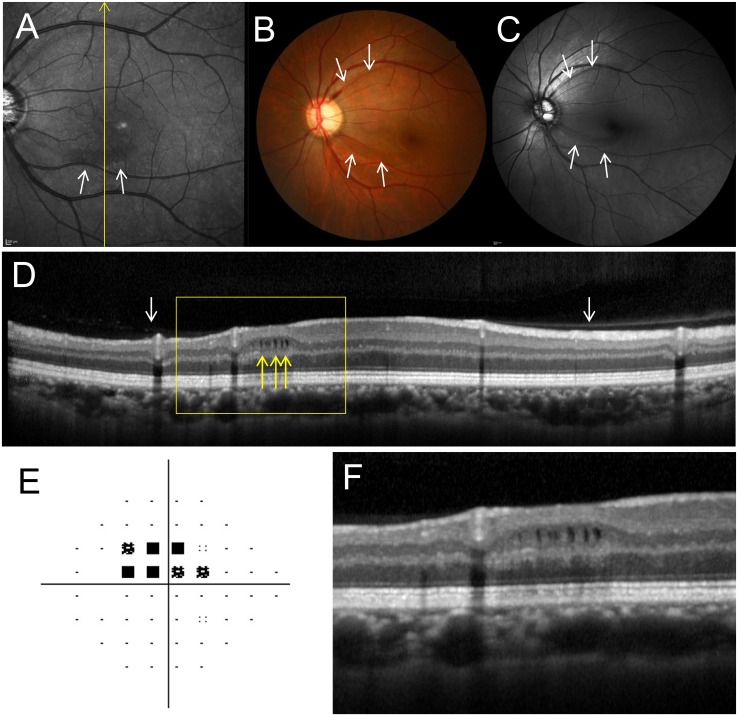
Fig 3. The left fundus of a 44-year-old woman with primary open angle glaucoma and microcystic inner nuclear layer (INL) lesions.
A, Infrared image shows perimacular hyporeflective patterns in the region with INL microcystic lesions (white arrows). B, C, Fundus (B) and red-free (C) photographs show retinal nerve fiber layer defects (NFLD, white arrows). Disc hemorrhage was also present at the upper NFLD. D, A Spectralis optical coherence tomography (OCT) image along the yellow line in A shows microcystic INL lesions (yellow arrows). The INL is thicker and the retinal nerve fiber and ganglion cell layers are thinner in the microcystic lesion area. This eye had a partial posterior vitreous detachment (white arrows). E, Pattern deviation map from Humphrey Visual Field Analyzer testing (24–2 Swedish interactive threshold algorithm standard program) showing visual field defects. An absolute scotoma at the superonasal test points closest to fixation corresponds to the location of the lower NFLD and microcystic INL lesions. F, Magnified OCT image (yellow box in D).

