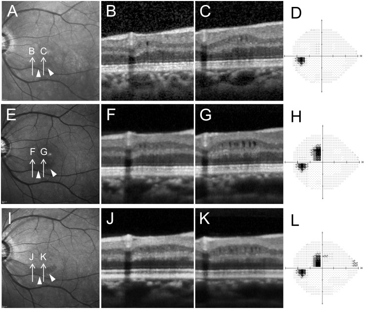
Fig 5. Changes over time in an eye with microcystic inner nuclear layer (INL).
Changes appearing before significant visual field damage occurred. A-D, E-H and I-L show images and testing obtained in December of 2008, 2012, and 2013, respectively. A, E, I, Infrared images showed perimacular hyporeflective patterns (arrow heads) becoming more obvious over time. B, C, F, G, J, K, Spectralis OCT images oriented along arrows in A, E and I. D, H and L, Standard automated perimetry testing results (Humphrey Visual Field Analyzer, 24–2 Swedish interactive threshold algorithm standard program gray scale). Subtle microcystic changes were observed in INL (B, C). It was noticed that localized thinning of retinal nerve fiber layer and ganglion cell layer existed though no severe visual field defects were present (B, C and D). Microcystic changes became more distinct as visual field defects progressed (F, G and H). Microcystic lesion changes became less apparent (F, G, J and K) while the visual field remained stable (H, L).

