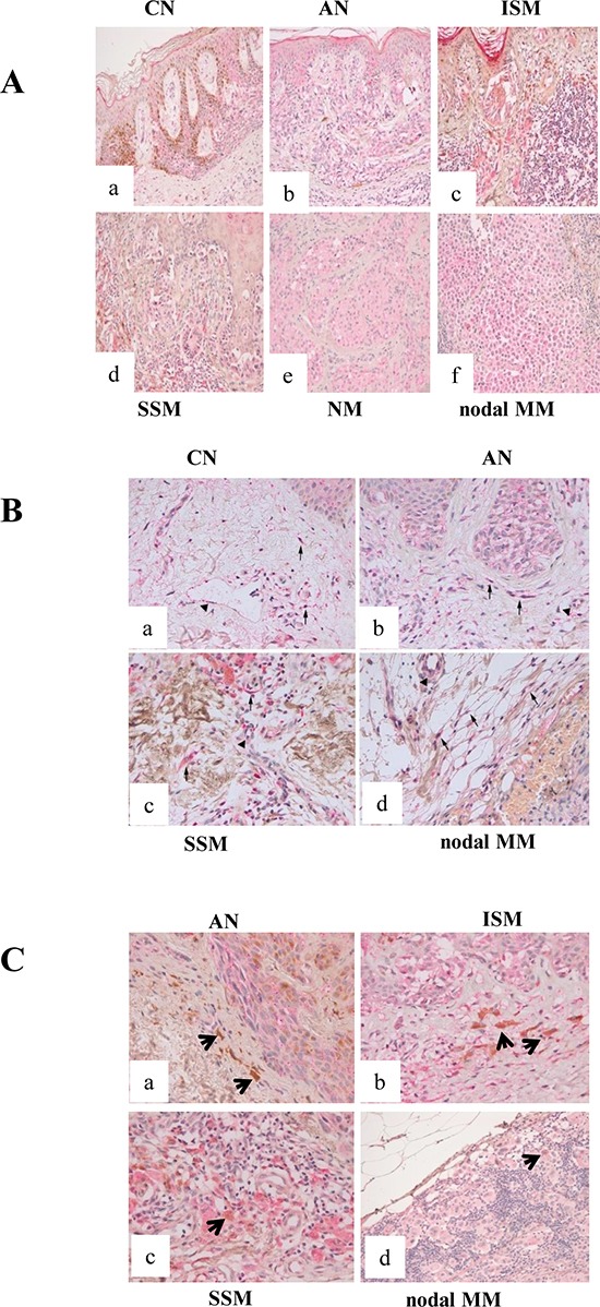
Figure 1. β3-ARs expression in human samples.

(A) β3-ARs expression in human cutaneous melanocytic lesions (X200). β3-AR immunostaining in junctional CN (a), AN (b), ISM (c), SSM (d), NM (e), nodal MM (f): all lesions show a low reaction intensity, confined to the cell cytoplasm. Epidermal keratinocytes exhibit staining, with strong positivity in granular layer; stromal cells also show reactivity. (B) β3-ARs expression in the microenvironment of human cutaneous melanocytic lesions (X400). (a) normal skin adjacent to CN; (b) AN; (c) SSM; (d) nodal MM. Arrows point to positive fibroblasts and arrow-heads point to positive blood vessels. Epidermis and in particular granular layer show positive reaction. Benign and malignant melanocytes are also stained. (C) Expression of β3-ARs in the microenvironment of human cutaneous melanocytic lesions. (a) AN and (b) ISM show melanophages (arrows) negative for β3-AR. (c) SSM and (d) nodal MM exhibit macrophages positive for β3-AR staining. Benign and malignant melanocytes are stained.
