
Figure 2. β-ARs expression in melanoma cells under various microenvironmental conditions.
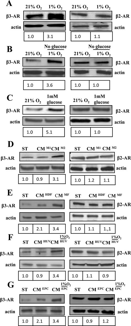
(A-G) 1 × 10⁶ A375 human melanoma cells were serum starved for 24 h and subsequently treated as reported in figure for 24 h. β3 and β2 ARs expression was evaluated in total lysates by immunoblotting analysis. Cells were exposed to (A) hypoxia (1%O2); (B) ischemia (1%O2 and 0% glucose); (C) hypoglycemia (1 mM) or exposed to CM derived from different activated stromal cells of tumor microenvironment: (D) M1 or M2 macrophages; (E) HDFs stimulated with TGFβ (myofibroblasts, MFs), as previously reported [55] or (F, G). HUVECs and EPCs exposed to hypoxic conditions. Anti-actin immunoblot was used to ensure equal protein loading. The figure is representative of three independent experiments.