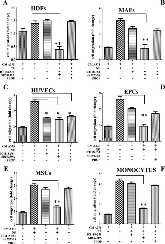
Figure 3. Recruitment of stromal cells.

Different stromal cells were allowed to migrate for 24 h toward CM derived from A375 melanoma cells. In left panels, A375 cells were incubated in the presence or absence of NE (10 μM) and/or β-ARs antagonist ICI 118-551 (1 μM), SR59230A (10 μM) and propranolol (1 μM). Figure shows recruitment of HDFs (A); MAFs (B); HUVECs (C); EPCs (D); MSCs (E) and monocytes (F). The figure is representative of three independent experiments. *P < 0.05, **P < 0.001, ***P < 0.0001 vs NE stimulated cells.
