
Figure 7. Cancer stemness induced by NE and HDFs.
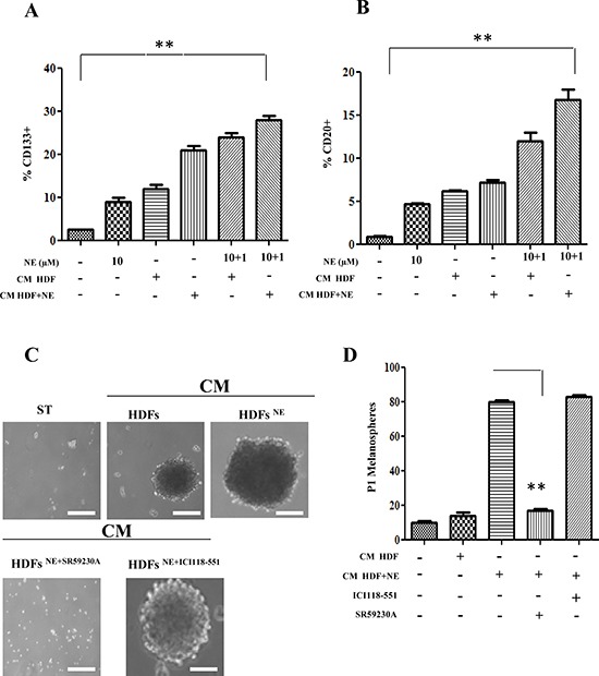
(A, B) Serum-starved A375 cells were stimulated for 24 h with CM derived from HDFs, incubated in the presence or absence of NE (10 μM) and/or β-ARs antagonist ICI 118-551 (1 μM), SR 59230A (10 μM). The last two samples were exposed again to NE (1 μM) for additional 24 h. Stem cells marker CD133 (A) and CD20 (B) were analysed by cytofluorimetric analysis **P < 0.001 vs NE stimulated cells. (C, D) A375 cells were serum starved for 24 h and incubated with CM derived from HDFs, preincubated with βARs antagonist ICI 118 (1 μM), SR59230A (10 μM). Cells were incubated in low-attachment dishes to evaluate melanospheres formation. Scale bars represent 100 μm. (C) After 10 days, pictures of P0 melanospheres were taken and representative images are shown. (D), P1 individual spheres derived after 10–15 days cultured cells, dissociated from single P0 melanospheres, were counted. Bar graphs show the mean ± SD of three independent experiments **P < 0.001 vs CM from HDFs-NE stimulated.