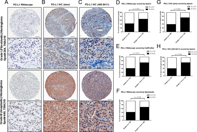
Figure 4. PD-L1 mRNA and protein expression in meningioma by grade in validation cohort.
A, Representative images of PD-L1 RNAscope staining, B, PD-L1 IHC (Sinobiological) antibody staining and C, PD-L1 IHC (405.9A11) antibody staining from a WHO grade I (fibroblastic) meningioma and from a WHO grade III (anaplastic) meningioma from a validation TMA (TMA310). Scale bar in low magnification, 100um; scale bar in high magnification, 20 um. PD-L1 RNAscope data was analyzed by D, Aperio and E, CellProfiler and F, Spotstudio™. Aperio pixel scoring was used to score PD-L1 IHC with the G, (Sinobiological) antibody and with the H, (405.9A11) antibody.

