
Fig. 2. Expression of CXCR4 and CXCL12 in pancreatic cancer tissues.
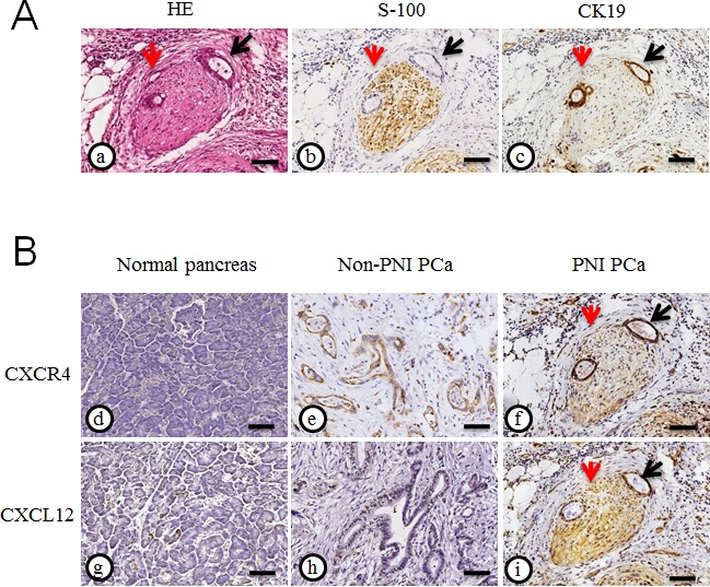
(A) HE staining (a), immunohistochemical staining of S100 (b) served as a nerve tissue marker and CK19 (c) served as a cancer cell marker in PCa tissues with PNI; (B) The representative immunohistochemical staining for CXCR4 and CXCL12 in the normal pancreas (d and g) and the resected PCa specimens accompanied without PNI (e and h) or with PNI (f and i) (200× magnification); the peripheral nerve (red arrow) was infiltrated by PCa cells (black arrow). Scale bar, 100μm.