
Figure 4. Immunohistochemical detection of brachyury protein in human lung cancers using MAb 54-1.
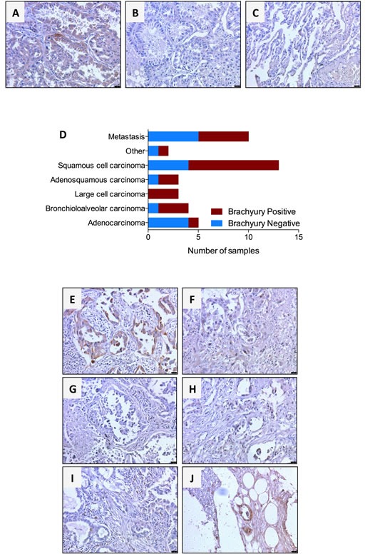
Transmitted light photomicrographs of a primary bronchioloalveolar carcinoma stained with (A) MAb 54-1 versus (B) control isotype IgG. Also shown is a representative staining of normal lung (C) with MAb 54-1. Expression of brachyury was analyzed by immunohistochemistry with MAb 54-1 in 30 cases of primary lung cancer and 10 lung cancer metastases. Shown (D) is the number of brachyury positive and brachyury negative cases for each tumor type. (E-J) Transmitted light photomicrographs of representative primary bronchioloalveolar (E) and large cell (F) primary lung carcinomas. Also shown are matched pairs of primary adenocarcinoma (G) and its corresponding bone metastasis (H) and a primary adenosquamous carcinoma (I) and corresponding matched bone metastasis (J). The brown signal corresponds to brachyury. Magnification 20X, scale bars = 100 μm.