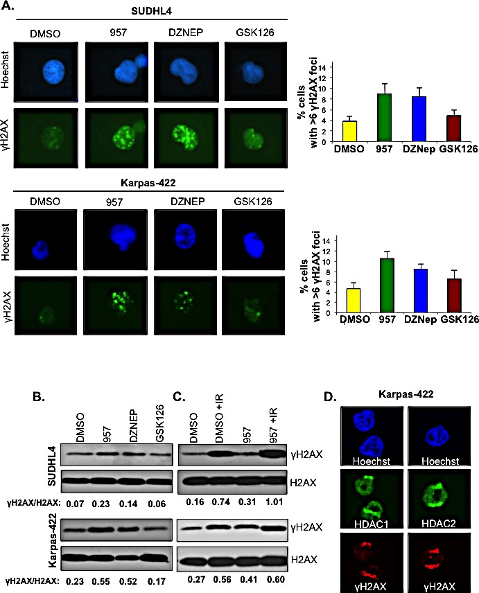
Figure 5. Selective inhibition of HDAC1,2 activates DNA damage response and impairs DSB repair in EZH2 DLBCL cells.

A: SUDHL4 and Karpas-422 cells were treated with DMSO, 2μM ACY-957, 0.5μM DZNEP or 0.5μM GSK126 for 48h and immunofluorescence staining with γH2AX was performed. The percentage of cells with 6 or greater γH2AX foci were counted in three independent experiments and at least 100 cells were counted in each experiment. The average with standard errors calculated from three independent experiments is shown in the figure. B: SUDHL4 and Karpas-422 cells were treated with DMSO, 2μM ACY-957, 0,5μM DZNEP or 0.5μM GSK126 for 48 h. Histones were purified using trichloroacetic acid extraction protocol (see methods). Western blot analysis was done with anti-γH2AX and histone H2AX served as the loading control. C: SUDHL4 and Karpas-422 cells were treated with DMSO or 2μM ACY-957 for 48h. Following DMSO or ACY-957 treatment, cells were exposed to a 5Gy dose of ionizing radiation and allowed to recover for 30 minutes prior to chromatin extraction. Western blot analysis was done with anti-γH2AX where total histone H2AX served as a loading control. D: Karpas-422 cells were micro-irradiated with laser and allowed to recover for 15 minutes before fixation and immunofluorescence staining with anti-γH2AX, anti-HDAC1 and anti-HDAC2 antibodies was done.
