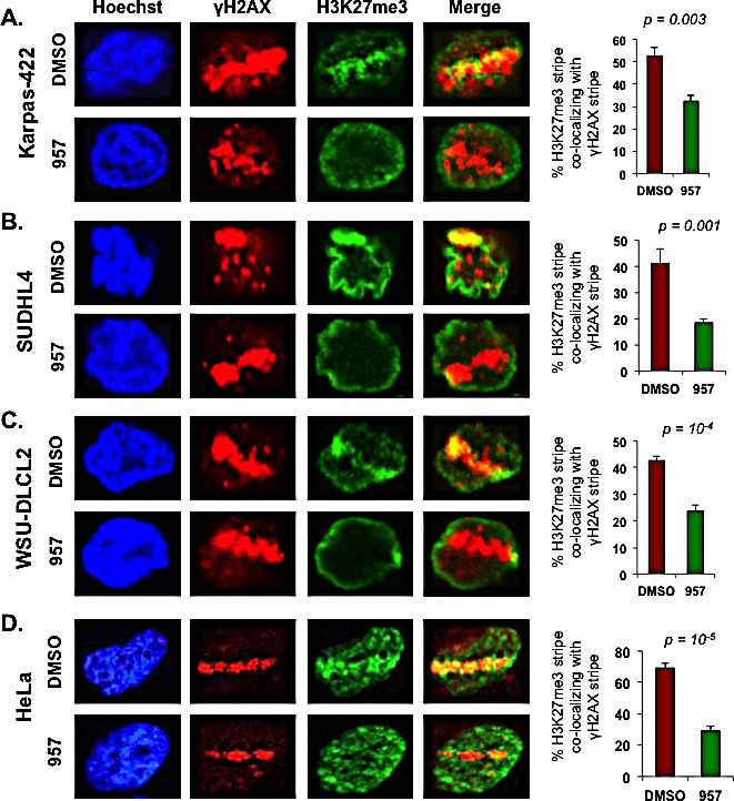
Figure 6. HDAC1,2 activity is critical for H3K27me3 enrichment at defined laser-induced break sites in chemoresistant DLBCL cells.

Karpas-422, SUDHL4, WSU-DLCL2 and HeLa cells were laser micro-irradiated and allowed to recover for 15 minutes before fixation and immunofluorescence staining with anti-γH2AX and anti-H3K27me3. Subsequently, the percentage of cells with H3K27me3 lines that co-localized with γH2AX were counted. At least 100 cells with γH2AX lines were counted in each experiment. The quantitation shown is the average calculated from independent experiments +/− standard error. Quantitation from five, seven, five and six independent experiments performed in Karpas-422, SUDHL4, WSU-DLCL2 and HeLa cells, respectively, were used in the graphs shown in this figure. Statistical analysis was performed and the p-values calculated from the t-test are shown in the figure. Merge is the overlay of γH2AX and H3K27me3 pictures.
