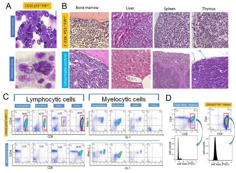
Figure 3. Histologic features and flow cytometry analysis of FIR+/−P53−/−.
(A) Atypical cells were indicated by Giemsa stain in bone marrow and peripheral blood of FIR+/−P53−/− mouse (C610). (B) Histologic features of bone marrow, liver, spleen and thymus in FIR+/−P53−/− (F338) and wild mouse by Hematoxylin-Eosin stain. (C) Flow cytometry analysis of lymphocytic cells with CD4 and CD8 as indicated markers. Mac1 and Gr1 were used for myelocytic markers. Flow cytometry analysis revealed that lymphocytic atypical cells (left) were CD4low+CD8+ phenotype (gated area) but no significant findings in myeloid cells (right) in FIR+/−P53−/− mouse (C610), and diagnosed as T-cell type acute lymphocytic/lymphoblastic leukemia (T-ALL)/lymphoma. (D) Cell size of gated area was measured by flow cytometry analysis (FSC: Forward Scatter).

