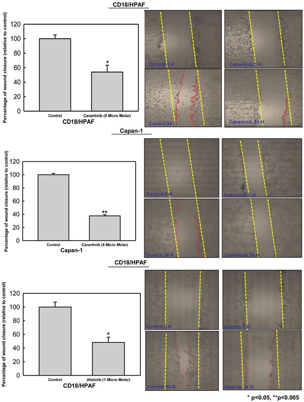
Figure 5. Canertinib and afatinib inhibits motility of pancreatic cancer cells.

CD18/HPAF and Capan-1 cells were trypsinized, counted and seeded at a density of 2×10⁶ cells in 60-mm Petri dishes and kept in 10% DMEM overnight. To determine the effect of canertinib and afatinib upon wound closure artificial wounds were created in 90% confluent cells and after 24 hour the cells were treated with vehicle DMSO (0.01%), canertinib 5 μM and afatinib 1 μM in complete medium. Images were taken at 0 and 24 hour in both control and inhibitor treated cells and cells migrated in the wound were measured by measuring the distance of migration The bars represents the percentage of wound closure between the control and treatment with significant p value less than 0.05 in CD18/HPAF (for both inhibitors) and p value less than 0.005 in capan-1 cells. Representative images of wound areas obtained at 0 and 24 hours before and after addition of canertinib and afatinib in pancreatic cancer cells. The red dotted lines depict the area of wound closure between the treatment and control cells.
