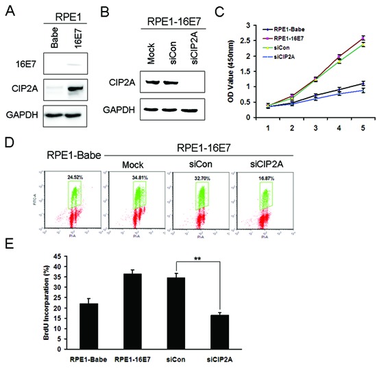
Figure 3. Knockdown of CIP2A inhibited cell proliferation and DNA synthesis of HPV-16E7-expressing cells.

(A) Western blot analysis of protein level of 16E7 and CIP2A in RPE1-16E7 cells and (B) with CIP2A siRNA for 48 hr. (C) CCK8 assay of cell proliferation of RPE1-16E7 cells with CIP2A siRNA. (D) Flow cytometry of cells with CIP2A siRNA and labeled with BrdU for 2 hr, then stained with PI and BrdU; and (E), Quantification. Babe, vector control. **, P < 0.01.
