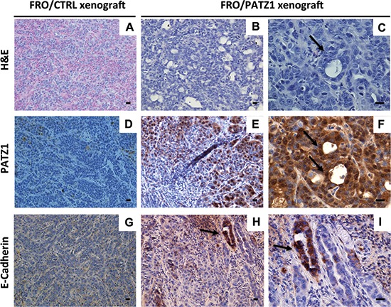
Figure 6. FRO/PATZ1 xenografts showed an epithelial-like phenotype.

Representative images of tumor tissues derived from nude mice injected with FRO/PATZ1 (B, C, E, F, H, I) or control (CTRL) cells (A, D, G) from the experiments shown in Figure 5C. Tumors developed from PATZ1-expressing cells showed features of epithelial-like differentiation represented by follicular-like structures (arrows). Consistently, E-cadherin immunostaining revealed a strong positive reaction in such follicular-like structures (H, I). Immunostaining for PATZ1 showed strong expression of PATZ protein in delimited areas including cells with an epithelial-like phenotype (E, F), whereas it was negative in all the other areas of PATZ1 xenografts (E) and in CTRL counterparts (D) Scale bars = 100 nm. H&E = hematoxilin and eosin staining.
