
Figure 7. IKK inhibition reduced growth of V2D1 tumors.
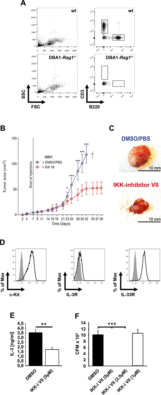
(A) Blood from wt or DBA1-Rag1−/−-mice was analyzed for CD3 and B220. (B) V2D1-cells (1 × 10⁶) were injected subcutaneously into the flanks of DBA/1-Rag1−/−-mice and tumor area was assessed for 6 weeks using a Mitutoyo Quick Mini caliper. Growing tumors were either treated with DMSO/PBS (blue line) or with IKK-inhibitor VII (red line) (25 μM). [Data represent the mean SD of 10 mice with tumors (p < 0,001)]. (C) V2D1 tumor size after 5 weeks, (upper panel) treated with vehicle DMSO/PBS and (lower panel) treated with IKK-inhibitor VII. (D) Cells from an explanted tumor were analyzed for surface expression of c-Kit, IL-3Rα and IL-33R. (E) Explanted cells were left untreated or were treated with the IKK-inhibitor VII. Supernatants were collected and analyzed for IL-3 (p < 0,01). (F) Explanted cells were treated with the IKK-inhibitor VII, were probed with [H3]thymidine and analyzed by β-counting (p < 0,001).