. 2015 Jun 15;4:e05061. doi: 10.7554/eLife.05061

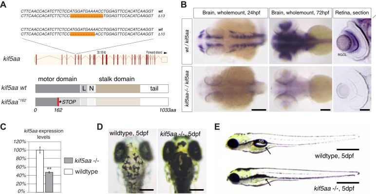
Figure 1. Generation of loss-of-function alleles of the anterograde motor protein Kif5aa.

(A) Employing TALENs targeting exon4 of the kif5aa open reading frame, we generated two loss-of-function alleles with a 10 bp and 13 bp deletion, respectively. These result in a frameshift at amino acid 122 and a premature stop codon after 162 of 1033aas within the motor domain of Kif5aa. L = linker region, N = neck region. (B) In situ hybridization shows a substantial downregulation of kif5aa mRNA in 24 hpf and 72 hpf old embryos. Scale bars (from left to right) = 150 μm, 100 μm, 50 μm. RGCL = Retinal Ganglion Cell layer. (C) Quantitative reverse transcription PCR confirms that only 47% of wild-type kif5aa mRNA expression levels are reached in homozygote mutant embryos at 4 dpf (p < 0.01). (D) Kif5aa mutant embryos show expanded melanosomes within their melanocytes and appear dark compared to wild-type embryos. Scale bars = 200 μm. (E) They fail to inflate their swim bladder and die 10 days post fertilization. Scale bars = 400 μm. Arrow: pointing at the respective location of the swim bladder. SB = swim bladder.

DOI: http://dx.doi.org/10.7554/eLife.05061.003

Figure 1.

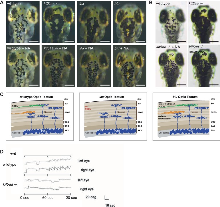
Figure 1—figure supplement 1. Melanosomes transport is not abolished in kif5aa mutants but they show no optokinetic response.

Figure 1—figure supplement 1.

(A) Phenotype of wild-type, kif5aa*162 mutant, lakritz and blumenkohl embryos at 5 dpf. All three mutants show expanded melanosomes and appear dark. Application of norepinephrine (NA) results in aggregation of melanosomes. Scale bar = 200 μm. (B) Application of NA leads to aggregation of melanosomes in kif5aa mutant embryos; washing out of the drug results in their re-expansion. Scale bar = 200 μm. (C) Schematics illustrating the organization of the optic tectum in wild-type, lakritz and blumenkohl embryos. The tectum is subdivided in multiple sublaminae. Retinal Ganglion Cells (RGC) axonal arbors grow into distinct layers within the tectal neuropil where they form functional synaptic connections with dendrites of periventricular neurons (PVNs). Lakritz mutant embryos lack all retinal input as they fail to specify RGCs. Blumenkohl mutants grow RGCs with increased axonal arbor sizes. The rate of synaptic transmission between RGCs and periventricular neurons is reduced. SO = stratum opticum, SFGS = stratum fibrosum et griseum superficiale, SGC = stratum griseum central, SAC = stratum album central, SPV = stratum periventriculare. (D) Kif5aa*162 mutant embryos do not show an optokinetic response. Eye positions (angles relative to a horizontal axis) were plotted over time during optokinetic stimulation in one direction. During the first 60 s, no stimulus is shown followed by 60 s of motion stimulation. The OKR has a sawtooth profile in siblings, consisting of alternating quick and slow phases while kif5aa*162 mutants do not show any response to the stimulus (n = 6).