. 2015 Jun 15;4:e05061. doi: 10.7554/eLife.05061

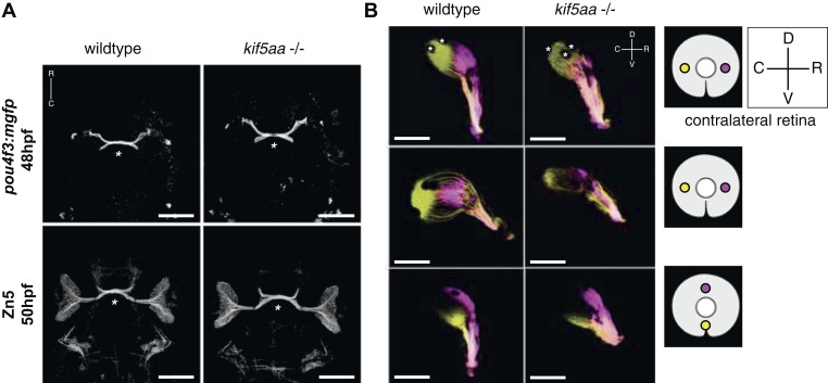
Figure 2. Outgrowth of the optic nerve and retinotopic mapping is normal in kif5aa mutants.

(A) Confocal imaging of the Tg(pou4f3:mGFP) transgene, labeling a subpopulation of Retinal Ganglion Cells (RGCs) with membrane bound GFP, at 48 hpf reveals that outgrowth of the optic nerve formed by RGC axons from the retina is not affected by the kif5aa mutation. Immunostaining against the Zn5 antigen (DM- GRASP/neurolin present within the visual system only on RGCs [Laessing and Stuermer, 1996; Fashena and Westerfield, 1999]) confirms that optic chiasm formation is normal (marked with an asterisk). No pathfinding errors occur at this level of axonal growth. Scale bars = 200 μm. Embryos facing upwards. R = rostral, C = caudal. (B) Injections of the lipophilic dyes DiI and DiO in different quadrants of the contralateral retina (depicted in the right panel) show that retinotopic mapping to the optic tectum is performed in the correct manner. Asterisks = pigment cells in the skin. D = dorsal, V = ventral, R = rostral, C = caudal. No misrouting of RGC axons to the ipsilateral tectum was observed (data not shown). Scale bars = 150 μm.

DOI: http://dx.doi.org/10.7554/eLife.05061.005

Figure 2.

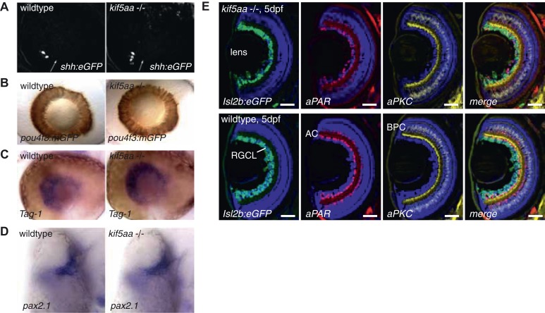
Figure 2—figure supplement 1. Patterning of the mutant retina and neurogenesis is not affected in mutants.

Figure 2—figure supplement 1.

(A) The expression of the Tg(shh:eGFP) transgene marks the onset of neurogenesis (white arrow) in the developing retina (Shkumatava et al., 2004) and its expression is not altered in kif5aa mutant retinae. (B) RGC differentiation marked by Tg(pou4f3:mGFP) (Xiao et al., 2005) expression is not affected. (C) The glycosylphosphatidyl inositol (GPI)-anchored protein of the immunoglobulin (Ig) superfamily Tag-1 I is expressed in nasal RGCs (Lang et al., 2001). In situ staining shows that nasal patterning is identical in wild-type and kif5aa mutant retinae. (D) Pax2.1, whose expression is restricted to the optic stalk and retinal cells around the choroid fissure and that mediates optic stalk and chiasm formation in fish (Krauss et al., 1991; Puschel et al., 1992) shows a normal expression pattern in kif5aa mutants. (E) Cryosection of Tg(Isl2:Gal4, UAS:eGFP) transgenic wild-type and mutant retinae and immunostaining against eGFP (RGCs) (Ben Fredj et al., 2010), Parvalbumin (marking amacrine cells) (Godinho et al., 2005) and protein kinase C (marking bipolar cells) (Godinho et al., 2005) shows that all cell types are present in the right retinal layer. Blue = DAPI. Scale bar = 10 μm. RGCL = RGC layer, AC = amacrine cells, BPC = bipolar cells.