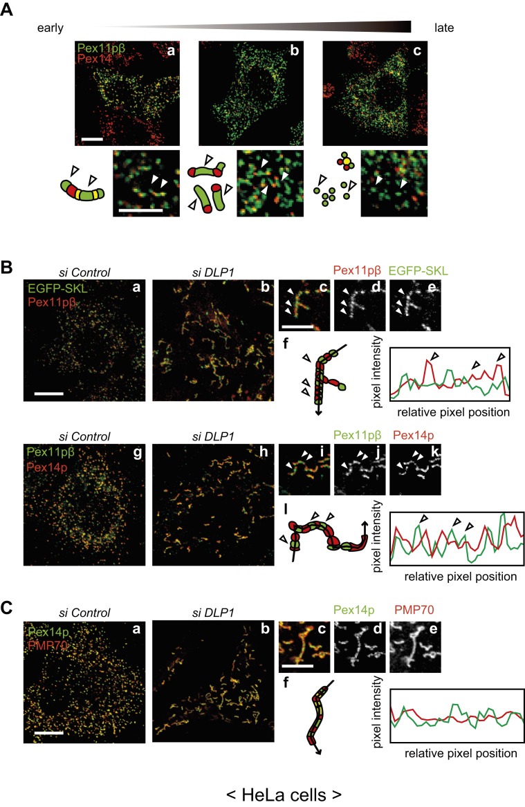
Fig. 1. Pex11pβ forms Pex11pβ-enriched membrane subregions on the peroxisomal membrane.
(A) HeLa cells were transfected with a plasmid encoding human Pex11pβ for 6–24 h and immunostained with antibodies against Pex11pβ (green) and Pex14p (red). Panels are arranged according to the stage of peroxisomal proliferation. Arrowheads indicate regions enriched in Pex11pβ compared with Pex14p. Scale bars: 10 µm and 5 µm (enlarged). (B) HeLa cells stably expressing EGFP-SKL (a–e) and control HeLa cells (g–k) were treated with a control siRNA (a,g) or DLP1 siRNA (b–e,h–k) for 72 h. Cells were subjected to immunostaining using the anti-Pex11pβ antibody (a–e) or double-stained with the anti-Pex11pβ antibody and an anti-Pex14p antibody (g–k). Magnified view of DLP1 siRNA-treated cells (c–e,i–k), and pixel intensity by line scanning along the elongated peroxisome (arrow) were plotted (f,l). Clear peaks of Pex11pβ were highlighted by arrowheads. Scale bars: 10 µm and 5 µm (enlarged). (C) HeLa cells were treated with a control siRNA (a) or DLP1 siRNA (b) for 72 h, and stained the DLP1 siRNA-treated cells with antibodies to Pex14p and PMP70 in magnified views (c–e). Line scanning of elongated peroxisomes was shown in (f). Scale bars: 10 µm and 5 µm (magnified view).

