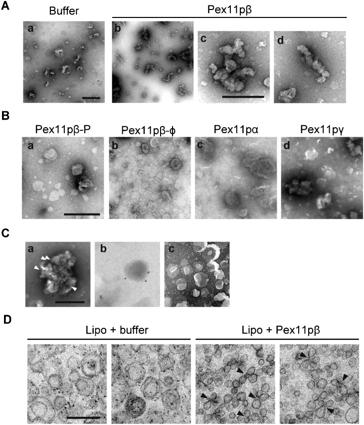
Fig. 6. Morphology of Pex11pβ-reconstituted proteo-liposomes.
(A–C) Negative staining electron micrographs of Pex11pβ-reconstituted proteo-liposomes. (A) Control liposomes (a) and Pex11pβ-reconstituted proteo-liposomes (b–d) were negatively stained with 2% uranyl acetate. Pex11pβ-reconstituted liposomes are magnified in panels c and d. Scale bars: 1 µm (a,b) and 0.5 µm (c,d). (B) Negative staining of proteo-liposomes reconstituted with Pex11pβ-P (a), Pex11pβ-Φ (b), Pex11pα (c), or Pex11pγ (d). Scale bar: 1 µm. (C) Reconstituted proteo-liposomes were subjected to immune labeling, and stained with uranyl acetate. Pex11pβ- (a) or Pex11pβ-P- (b) reconstituted proteo-liposomes were sequentially incubated with the anti-Pex11pβ antibody and an anti-rabbit IgG conjugated to 10 nm gold particles. The anti-Pex11pβ antibody and gold-labeled anti-rabbit IgG did not react with the control liposomes (c). Scale bar: 0.5 µm. Arrowheads indicate the gold particles (see text). (D) The reconstituted proteo-liposomes were subjected to electron microscopy. Control-treated liposomes (left) and Pex11pβ-reconstituted liposomes (right) were pelleted by ultracentrifugation and prepared for embedding and ultra-thin sectioning as described in the Materials and Methods. Note that the constriction of the liposomal membrane was observed in Pex11pβ-reconstituted liposomes (arrowheads). Scale bar: 0.2 µm.

