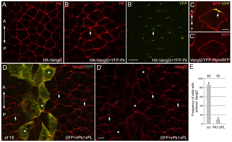
Fig. 3. Vangl2 polarity is directed by Prickle.
Early embryos were injected with HA-Vangl2 (150 pg) and YFP-Pk RNAs (100 pg) either separately or together as indicated. At early neural plate stage, embryos were fixed and immunostaned with anti-HA or anti-GFP antibodies. (A) HA-Vangl2 is homogeneously distributed at cell boundaries. (B,B′) The complex of Vangl2/Pk is polarized at the anterior end of each cell. HA-Vangl2 (B) and YFP-Pk (B′). (C,C′) The anterior localization of the Vangl2/YFP-Prickle complex in an expressing cell is visualized by YFP and membrane RFP (mRFP) epifluorescence. (D,E) Early embryos were coinjected with zPk1ΔPL RNA (1.5 to 2 ng) and GFP as lineage tracer (100 pg). (D,D′) Immunofluorescence reveals Vangl2 anterior accumulation (arrows) in the neural plate, with the exception of the cells containing zPk1ΔPL (marked by GFP, asterisks). Dorsal view of the neural plate midline is shown, anterior is to the top. (E) Quantitation of the data shown in D. See also Fig. 4C for control RNA effect. Error bars represent s.d. Co, control cells on the uninjected side. Scale bars are 20 µm in B,D, and 5 µm in C.

