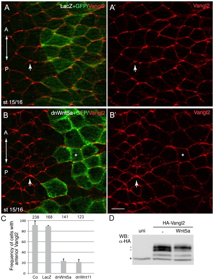
Fig. 4. A role of Wnt signaling in establishing the Vangl2 polarity.
(A,B) Early embryos were injected with DN-Wnt5a RNA (0.3 ng) and GFP RNA (0.1 ng) as a lineage tracer. Neural plate cells mosaically expressing this construct reveal lack of Vangl2 enrichment at the anterior of each cell (asterisks) as compared to non-expressing cells (arrows). A', B' are single-channel images corresponding to A and B. Control LacZ RNA (1.5 ng) coinjected with GFP RNA (0.1 ng) had no effect on anterior distribution of Vangl2. Dorsal view of the neural plate is shown, anterior is to the top. Scale bar, 20 µm. (C) Quantification of data from the experiments with DN-Wnt constructs showing mean frequencies of cells with anterior Vangl2±s.d. At least 5–10 embryos were examined per each treatment. Numbers of scored cells are shown on top of each bar. (D) Vangl2 is phosphorylated in response to Wnt5a in Xenopus ectoderm. Early embryos were injected with HA-Vangl2 mRNA (0.1 ng) together with Wnt5a RNA (0.5 ng). Cell lysates were prepared at the midgastrula stage (st. 11) for western analysis with anti-HA antibodies. Wnt5a causes HA-Vangl2 to migrate slower in whole embryo lysates (st. 11). Asterisk indicates a non-specific protein band reflecting protein loading.

