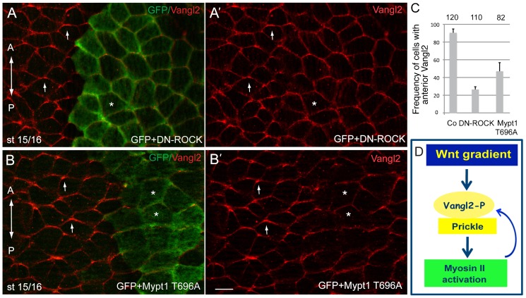
Fig. 6. Feedback regulation of AP-PCP by the ROCK/Myosin II pathway.
(A,B) Eight cell embryos were unilaterally coinjected with the lineage tracer (GFP RNA, 100 pg) and DN-ROCK RNA (100 pg, A) or Mypt1T696A RNA (100 pg, B) and Vangl2 polarity was assessed by immunostaining at the neural plate stage. Both the interference with ROCK signaling (A,A′) and the dephosphorylation of Myosin II light chain by the myosin phosphatase Mypt1 (B, B′) inhibit Vangl2 polarity. Arrows point to polarized Vangl2 staining. Asterisks indicate treated cells marked by GFP with non-polarized Vangl2. (C) Quantification of the data shown in A,B. Error bars represent s.d. (D) Model of AP-PCP. Wnt activity gradient leads to the polarization of the Vangl2/Pk complex to the anterior cell domain. Myosin II activity, a putative target of PCP signaling, mediates feedback regulation of core PCP protein localization. Scale bar, 20 µm.

