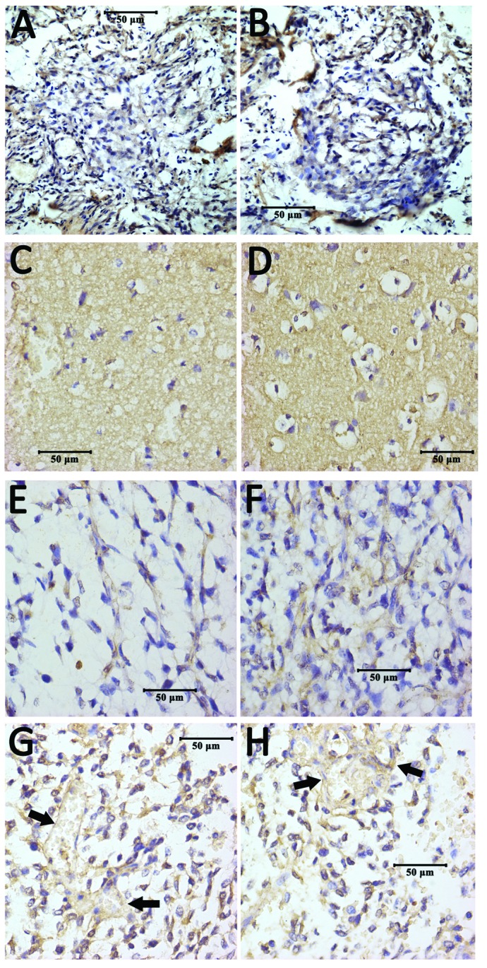
Figure 5.

Immunohistochemical staining of ADAM10 protein. (A and B) Representative images from the control group, in which there were no ADAM10 positive cells visible. (C and D) Images from the low-grade group, in which the structure of many tumor cells had been destroyed and the cells appeared vacuous with non-specific background staining; in (D), partial positive staining is observed, which is scattered in tumor cell membranes and in the cytoplasm. (E and F) Representative images from the high-grade group. A relatively greater number of tumor cells with positive staining are present in (E), which is even more apparent compared with (F), where positive staining is demonstrated to be further increased in the perinuclear membrane or the cytoplasm of tumor cells. (G and H) Images from high-grade gliomas, in which blood vessel walls display positive staining (arrows), and the nuclei are stained by hematoxylin. ADAM10, a-disintegrin and metalloproteinase 10.
