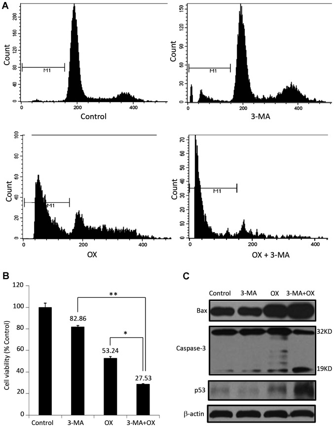
Figure 3.
3-MA enhances OX-induced apoptosis in CT26 cells in vitro. (A) Apoptosis analysis of CT26 cells by flow cytometry. The sub-G1 cell population percentage in OX treated cells was 39.48%; however, it was increased to 86.81% in 3-MA plus OX cotreated cells. (B) The viability of CT26 cells administered with different treatment regimens was measured by performing an MTT assay. *P<0.05; **P<0.01. (C) The expression of apoptotic proteins, such as Bax, caspase-3 and p53, was detected by performing a western blot analysis in CT26 cells administered with different treatment regimens. β-actin was used as the loading control. 3-MA, 3-methyladenine; OX, oxaliplatin; Bax, Bcl-2-associated X protein.

