Figure 1.
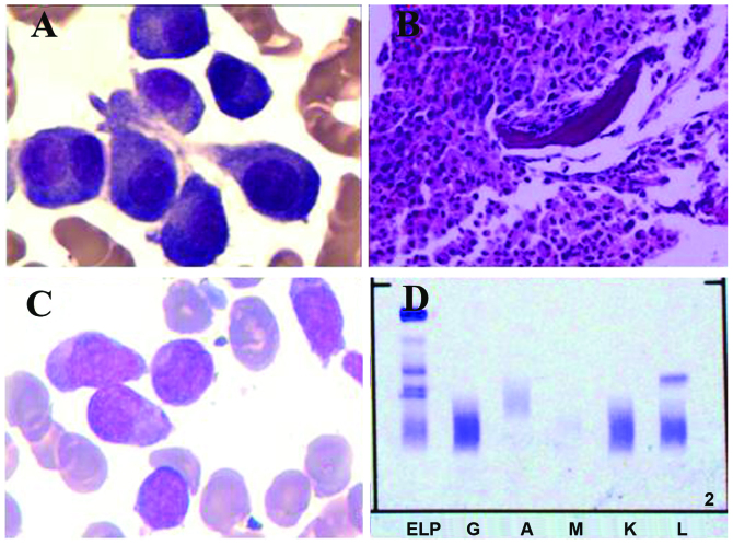
A bone marrow aspirate and biopsy were performed. (A) Bone marrow aspirate results showing a cluster of abnormal plasma cells, including multinucleated plasma cells. (B) Bone marrow biopsy analysis showing an accumulation of myeloid cells, dominated by band forms and segmented neutrophils. A number of clusters of middle-sized cells with abundant cytoplasm were identified and immunochemistry results were CD38(+), CD138(+) and epithelial membrane antigen(+/-). (C) Bone marrow smear results demonstrating hypercellularity, with monoblast cells and promonocytes accounting for 58% of nucleated cells, indicating that the patient had progressed to acute monocytic leukemia. (D) Serum protein immunofixation electrophoresis showing a sharp positive band for the λ-light chain subtype. CD, cluster of differentiation. ELP, electrophoresis of protein; G, IgG; A, IgA; M, IgM; K, κ light chain; L, λ light chain.

